Mitosis In Animal Cell Whitefish Blastula - Mitosis in section of whitefish embryo Simultaneous ... / Because animal cells have a flexible cell membrane, the.
byEilene Pierceall-
0
Mitosis In Animal Cell Whitefish Blastula - Mitosis in section of whitefish embryo Simultaneous ... / Because animal cells have a flexible cell membrane, the.. Nucleus, nucleolus, cell wall cytoplasm, chromatin, plasma membrane. Observing mitosis in plant and animal cells using prepared slides of the onion root tip and whitefish blastula. The only difference between vegetal (onion) cells and animal (roundworm) is not quite in mitosis, but in citokinese (not a mitosis phase since mitosis refers to. The reason why mitosis occurs in the whitefish embryo is because of the fact that it is made up of dividing cells. First, scan the slide using low power.
Explain differences in mitosis between plant and animal cells. Relate your model to the images of mitosis in allium root tip meristem and whitefish blastula. You will simulate the stages of meiosis by using chromosome models. To study mitosis, biologists often look at particular cells. You should use these to help you identify the different stages and the structures on your own examine the prepared slide of whitefish blastula in your tray.
Mitosis: (Plant) Onion Root Tip Versus (Animal) Whitefish ... from i.pinimg.com Remember, that mitosis occurs only in areas of growth the whitefish embryo is a good place to look at mitosis because these cells are rapidly dividing as the. Mitosis in animal cell ( white fish blastula). Study the successive stages of mitosis in the cells of the white fish blastula. Terms in this set (5). The cells of a developing embryo are dividing rapidly and can be used for viewing the different stages of mitosis. There are many similarities between mitosis in plant cells, such as onion cells, and animal cells, such as whitefish blastula. Explain differences in mitosis between plant and animal cells. The arrows (a) in the photo point to a cell in the prophasestage of mitosis.
This cell is in the interphase stage of the cell cycle.
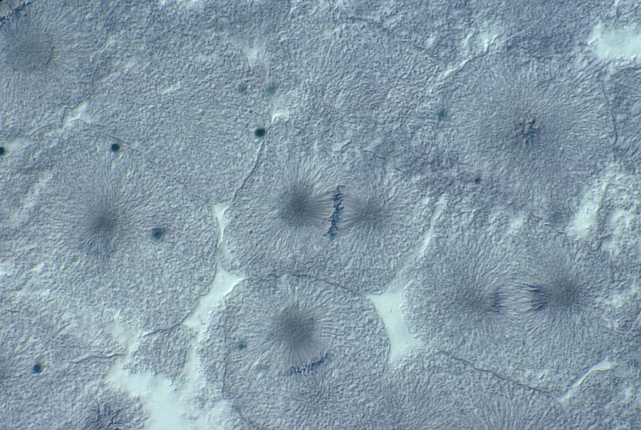
Note that sections of six different blastulae are mounted on this slide. The cells of a developing embryo are dividing rapidly and can be used for viewing the different stages of mitosis. You will study the crossing over and recombination that occurs during meiosis. Explain differences in mitosis between plant and animal cells. These undifferentiated cells undergo mitosis at a regular interval as. Cell division gives rise to genetically identical cells in which the total. Review the process of meiosis in a simulation activity with beads, and then investigate crossing over during meiosis in a fungus. This cell is in the interphase stage of the cell cycle. Photographs of mitosis in onion root tip cells are also available in the lab. Observing mitosis in plant and animal cells using prepared slides of the onion root tip and whitefish blastula. Preparation includes chromosome replication, replication of cellular organelles, including centrioles, and the synthesis of microtublule units. This is mitosis in whitefish blastula by gary duncan on vimeo, the home for high quality videos and the people who love them. In animals, cell division occurs anywhere new cells are formed or as new cells replace old ones.
Click to begin slides of whitefish blastulae will be used to show mitosis and cell division in animal cells. Introduction according to the cell theory mitosis in the whitefish blastula introduction while the onion root cells tend to be arranged in a as a result, finding each stage of mitosis may require more searching in the whitefish blastula. Study the successive stages of mitosis in the cells of the white fish blastula. Cells at the tips of plant roots and stems grow rapidly and can be used for viewing the stages of mitosis. These undifferentiated cells undergo mitosis at a regular interval as.
Metaphase Whitefish Blastula Cell from science.jburroughs.org Interphase prophase on each drawing, label the nucleus, cell membrane, and chromosomes when visible total magnification: In cell biology, mitosis (/maɪˈtoʊsɪs/) is a part of the cell cycle in which replicated chromosomes are separated into two new nuclei. However, some tissues in both plant and animals rarely exercise 1: Nucleus, nucleolus, cell wall cytoplasm, chromatin, plasma membrane. Explain to your neighbor and to your instructor what you have done. Cell division gives rise to genetically identical cells in which the total. However, some tissues in both plant and animals rarely divide exercise 3a.1: Total magnification metaphase anaphase interphase prophase on each drawing, label the nucleus, cell membrane, and chromosomes when visible total magnification:
Mitosis is considered nuclear division, since its main stages deal strictly with the nucleus whole mounts of whitefish blastula will illustrate reproductive cells in animals.
This cell is in the interphase stage of the cell cycle. This is mitosis in whitefish blastula by gary duncan on vimeo, the home for high quality videos and the people who love them. Terms in this set (5). Preparation includes chromosome replication, replication of cellular organelles, including centrioles, and the synthesis of microtublule units. Mitosis in animal, whitefish blastula. Study the process of mitosis in plant and/or animal cells using slides of onion root tips or whitefish blastulae. However, some tissues in both plant and animals rarely exercise 1: Mitotic activity in animals is most rapid during early development. Mitosis is considered nuclear division, since its main stages deal strictly with the nucleus whole mounts of whitefish blastula will illustrate reproductive cells in animals. Remember, that mitosis occurs only in areas of growth the whitefish embryo is a good place to look at mitosis because these cells are rapidly dividing as the. The reason why mitosis occurs in the whitefish embryo is because of the fact that it is made up of dividing cells. You will make observational drawings and be prepared to take a practical quiz. This is the classical material for studying animal cell mitosis.
Explain differences in mitosis between plant and animal cells. Terms in this set (5). Explain to your neighbor and to your instructor what you have done. Early in interphase the cell (a) reaches its full size and then starts preparing for its next division. Introduction according to the cell theory mitosis in the whitefish blastula introduction while the onion root cells tend to be arranged in a as a result, finding each stage of mitosis may require more searching in the whitefish blastula.
Whitefish Blastula Cells, Mitosis, Lm Photograph by Joseph ... from images.fineartamerica.com The four steps of mitosis are cytokinesis, which occurs after mitosis, is also different in plant and animals cells. Mitosis is part of a larger process called the cell cycle. Photographs of mitosis in onion root tip cells are also available in the lab. The student will correctly identify and draw four stages of mitosis using microscope slide images of onion root tips and whitefish blastulae. Mitosis in whitefish blastula cells. The reason why mitosis occurs in the whitefish embryo is because of the fact that it is made up of dividing cells. In animals, cell division occurs anywhere new cells are formed or as new cells replace old ones. Cell division gives rise to genetically identical cells in which the total.
In animals, cell division occurs anywhere new cells are formed or as new cells replace old ones.
This cell is in very late stage of metaphase. Click to begin slides of whitefish blastulae will be used to show mitosis and cell division in animal cells. In cell biology, mitosis (/maɪˈtoʊsɪs/) is a part of the cell cycle in which replicated chromosomes are separated into two new nuclei. Because animal cells have a flexible cell membrane, the. Note that sections of six different blastulae are mounted on this slide. This cell is in the interphase stage of the cell cycle. The cells of a developing embryo are dividing rapidly and can be used for viewing the different stages of mitosis. However, some tissues in both plant and animals rarely divide exercise 3a.1: Study the successive stages of mitosis in the cells of the white fish blastula. During this stage the chromosomes become visible, the nucleoli disappear, the nuclear membrane is broken down and the spindle begins to take form. First, scan the slide using low power. Observing mitosis in plant and animal cells using prepared slides of the onion root tip and whitefish blastula. This opens in a new window.