Animal Cell In Light Microscope - Gudu Ngiseng Blog Animal Cell Light Microscope / The organelles in a cheek cell that are not visible under a light microscope are the ribosomes.
byEilene Pierceall-
0
Animal Cell In Light Microscope - Gudu Ngiseng Blog Animal Cell Light Microscope / The organelles in a cheek cell that are not visible under a light microscope are the ribosomes.. Two important factors in microscopy are magnification and resolving power. We say cells are microscopic because they can only be seen under a microscope. You can see a variety of cells pretty well with the light microscope. In this type of microscope, there are ocular lenses in the binocular eyepieces and objective lenses in a rotating nosepiece closer to the specimen. To even see a boundary clear would require a stain that soaks.
Visible light passes and is bent through the lens system to enable the user to see the. At the end of the activity, the student should be able to: Image:plant cell seen under electron microscope. Most of the cells size range between 1 and 100 micrometers and are visible only with the microscope. Light microscopy (the use of microscopes is called microscopy), in plant cells c.
71 714 Plant Cell Stock Photos Pictures Royalty Free Images Istock from media.istockphoto.com Light microscope uses the properties of light to produce an enlarged image. Light microscopes using visible light and lenses to form a magnified image of the object under investigation e.g. In fact, hooke coined the term cell, in a biological context, when he described the microscopic structure of cork like a tiny, bare room or. These organelles are responsible for protein synthesis. Glass lenses enlarge the image and project it into the human eye or camera. It is the simplest type of microscope. The two visible structures that are visible with 'light microscope' are the cell wall and the vacuoles. The animal cell is more fluid or elastic or malleable in structure;
The patterns of light and dark that are recognized as an image of the specimen.
First seen with light microscopy 2. Light microscope uses the properties of light to produce an enlarged image. The different part of a cell are called subcellular structures. With a light microscope you can see several structures inside the cell. As you how does a light microscope work? With a light microscope you can see individual cells and large subcellular structures like the nucleus, but not internal cell structures such as. Use electromagnets to focus electrons resulting in significantly greater magnifications and resolutions. A cell is the smallest functional and structural entity of life that it is easier observing animal cell under light microscope. This is made of cellulose and is very rigid. Visible light passes and is bent through the lens system to enable the user to see the. Animal cell features (light microscope). Most cells are very small, and their structures can only be seen by using a microscope. Learn about and revise cells in animals and plants with this bbc bitesize combined science aqa synergy study guide.
Cells consist of cytoplasm enclosed within a membrane, which contains many biomolecules such as proteins and nucleic acids.2 most plant and animal cells are only visible under a light microscope, with dimensions between 1 and 100 micrometres.3 electron microscopy gives a much higher. It also has a very high resolving power. For example, both animal and plant cells are classified as eukaryotic cells, whereas bacterial cells are classified as prokaryotic. When you look at animal or plant cells under the electron microscope, you can see a lot more detail. As you how does a light microscope work?
File Animal Cell Optical Microscope View Jpg Wikimedia Commons from upload.wikimedia.org Light rays pass through the specimen on a slide. A compound light microscope is a microscope with more than one lens and its own light source. With the invention of the electron microscope a whole new world was open up to scientists. We say cells are microscopic because they can only be seen under a microscope. (2) study of animal cells : Resolving power is the ability to distinguish between separate things which are close to each other. Most cells are very small, and their structures can only be seen by using a microscope. In this type of microscope, there are ocular lenses in the binocular eyepieces and objective lenses in a rotating nosepiece closer to the specimen.
Cell ultrastructure and the importance of the cytoskeleton of cells.
Although sometimes found as monocular with one ocular. First seen with light microscopy 2. However, light microscopes form real colour images and can be used to watch living processes occur in microscopic detail, while electron microscopes cannot be used to study living cells. Under the light microscope, three parts can be seen in the animal cell: A compound light microscope is a microscope with more than one lens and its own light source. This is one of the tenets of the cell theory, a basic theory of biology. Learn about and revise cells in animals and plants with this bbc bitesize combined science aqa synergy study guide. This diagram shows a typical animal cell. Use electromagnets to focus electrons resulting in significantly greater magnifications and resolutions. We use microscope comprehensively in microbiology, mineralogy, cell biology, biotechnology, nano physics, microelectronics, pharmacology, and forensics. Light microscopy (the use of microscopes is called microscopy), in plant cells c. Light microscope uses the properties of light to produce an enlarged image. Vacuole,in plant cell it is very big in size but in animal cell they may appear but vry smallin sizecell wall,present only in plant cell.
Most light microscopes will enlarge a specimen up to 1000 times (1000x) but the electron. Visible light passes and is bent through the lens system to enable the user to see the. Two important factors in microscopy are magnification and resolving power. Cell ultrastructure and the importance of the cytoskeleton of cells. An iris diaphragm on the substage condenser controls the amount of light reaching the objective, and also affects the contrast of the specimen.
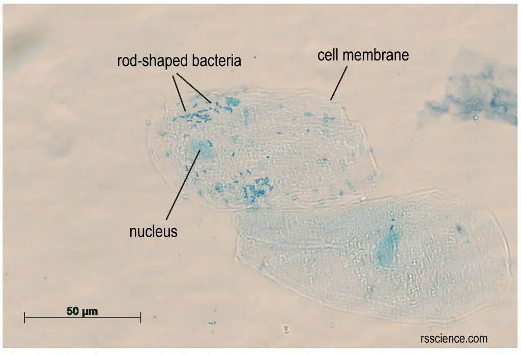
What Living Things You Can See Under A Light Microscope Rs Science from rsscience.com Most of the cells size range between 1 and 100 micrometers and are visible only with the microscope. An animation that shows animal cells. A cell is a very tiny structure which exists in living bodies. An electron microscope is required for virus and dna. In fact, hooke coined the term cell, in a biological context, when he described the microscopic structure of cork like a tiny, bare room or. A cell is the smallest functional and structural entity of life that it is easier observing animal cell under light microscope. Most commonly used microscopes are classified as light microscopes (figure 7.2a). Image:animal cell seen under light microscope.
However, light microscopes form real colour images and can be used to watch living processes occur in microscopic detail, while electron microscopes cannot be used to study living cells.
Most cells are visible under a light microscope, but mitochondria and bacteria are barely visible. Visible light passes and is bent through the lens system to enable the user to see the. Light microscope uses the properties of light to produce an enlarged image. This diagram shows a typical animal cell. It is meant for students with little to no experience preparing slides or using a compound light microscope. Level suitable for as biology. An electron microscope is required for virus and dna. These structures are discussed in more detail in the following pages. However, as you probably noticed in the previous activity. However, light microscopes form real colour images and can be used to watch living processes occur in microscopic detail, while electron microscopes cannot be used to study living cells. Although sometimes found as monocular with one ocular. It also has a very high resolving power. Animal cells are of various sizes and have irregular shapes.